 |
Témavezető | |
| E-mail cím |
|
|
| Kutatási terület | Application of nuclear physics and of accelerators | |
| Kutatásban résztvevők | ||
| Eszközök | ||
| Publikációk |
Three-dimensional imaging of surface chemical processes in catalysis can be carried out using high-resolution positron emission tomography.
The positron emission tomography (PET) technique is a well-known method in diagnostic nuclear medicine for the molecular imaging of biochemical functions in the human body. However, it can also be applied to the molecular imaging of chemical processes on the surfaces of heterogeneous catalysts. Dynamic studies of adsorption, desorption, and realignment of 11C positron-emitter labelled
compounds can be performed. Surface imaging techniques help one to look inside a catalyst bed, and understand the kinetics and surface dynamics of catalysis and the reaction mechanisms on surface sites under different experimental conditions.
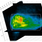
A small PET scanner (MiniPET-2) had been developed in Atomki in collaboration with the University of Debrecen, for small animal preclinical imaging (with parameters similar to those of a commercial small-animal PET apparatus). The camera has 12 detector modules in a full ring configuration. The 3D image is formed from 35 cross-sectional slices. Such PET scanners have great potential for both academic and industrial research in imaging catalyst surfaces, whether they are well- or poorly-functioning, or partially covered. The imaging system is powerful because the sample is scanned both radially and axially, so that the distribution of radioactive compounds is
analyzed in the total catalyst volume. This PET camera was used to map the location and quantitative distribution of a 11C -methanol compound in a zeolite catalyst bed (4 cm long and 1.6 cm across) in three dimensions.
 Magyar
